Home
/ Plant Cell Under Microscope Labeled - Microscope Onion Cell Labeled - Micropedia / The image resolution 800 x 708 px and the image size only 0 kb.
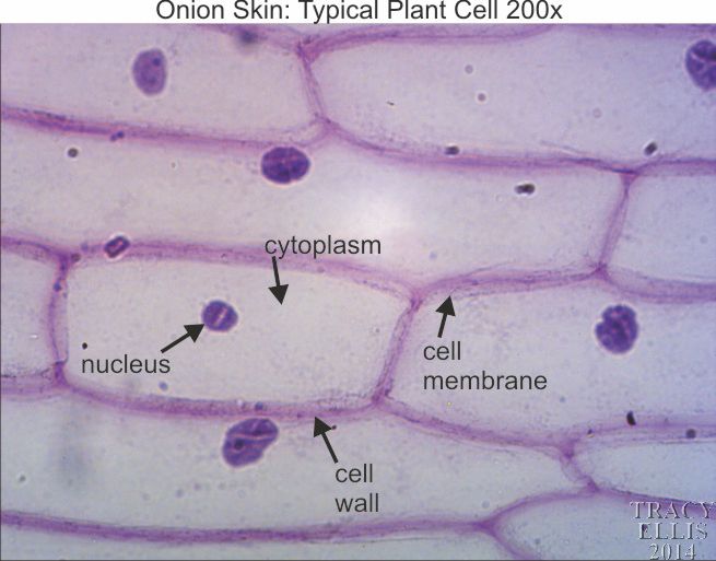
Plant Cell Under Microscope Labeled - Microscope Onion Cell Labeled - Micropedia / The image resolution 800 x 708 px and the image size only 0 kb.
Plant Cell Under Microscope Labeled - Microscope Onion Cell Labeled - Micropedia / The image resolution 800 x 708 px and the image size only 0 kb.. View plant cells under a microscope. Here's a diagram of a plant cell: Under a microscope, plant cells are surrounded by cell wall, which are not present in animal cells. (ii) presence of large central vacuole in plant cell. Here's a photo of a plant cell under an electron microscope.
Students will finish plant cell diagrams from monday. As you can see in the above labeled plant cell diagram under light microscope. In truth, there are still features of plant and animal cells we're only lately discovering. Labelled diagram of a plant cell under microscope posted on march 18 2011 by admin onion cells stained with methylene blue look at the images of onion cells as they would be seen under a microscope draw each magnification label appear high picture plant and animal cell … Given below is the diagram of a cell as seen under the microscope after having been placed in a solution
Plant Cell Under Microscope Labeled from brazile.education However, the internal structure and organelles are more or less similar. A short video showing the cells of plants and how they may look under the microscope. Learn vocabulary, terms and more with flashcards, games and other study tools. Plant structure and cross section botanical biology labeled diagrams collection. Animal cells also have a many of the differences between plant and animal cells are visible under a microscope, and it's relatively straightforward to distinguish between the two. A cell is a very tiny structure which exists in living bodies. Start studying plant cell labeling. Animal cells introduction background information:
Microscope slide cover slip onion.
We say cells are microscopic because they can only be seen under a microscope. 15) observe the elodea cells under the microscope, sketch one cell in the space provided below with the best magnification you can and label what you see. Lets get looking at some real plant cells! Structure of a plant cell. Start studying plant cell labeling. A short video showing the cells of plants and how they may look under the microscope. (ii) presence of large central vacuole in plant cell. See how a generalized structure of an animal cell and plant cell look with labeled diagrams. To learn how to get the best image from a microscope. They must draw and label the nucleus, cell continue with more related things as follows plant cell diagram without labels, microscope parts labeled and compound light microscope parts blank. The image resolution 800 x 708 px and the image size only 0 kb. In truth, there are still features of plant and animal cells we're only lately discovering. Labelled diagram of a plant cell under microscope posted on march 18 2011 by admin onion cells stained with methylene blue look at the images of onion cells as they would be seen under a microscope draw each magnification label appear high picture plant and animal cell …
Amazing pictures of 8 pictures of plant cells under a microscope is totally great for your biological science knowledge. As you can see in the above labeled plant cell diagram under light microscope. Observe the labeled diagram of plant cell. Lets get looking at some real plant cells! Here's a diagram of a plant cell:
Microscope Onion Cell Labeled - Micropedia from dissectionconnection.com.au Major differences between a plant cell and on animal cell are (i) presence of chloroplast in plant cell. The microscope consists of a stand (base + neck), on which is mounted the stage (for holding there are three structures that distinguish plant cells from animal cells. For immunofluorescence microscopy of plant cell walls, the first step consists in incubating the plant material in a fixative solution that commonly contains 4 once soaked in a solvent, the slides are covered with a cover slip and ready for inspection under microscope. Viewing the leaf under the microscope shows different types of cells that serve various functions. Plant and animal cells are similar, consisting of a protoplast bounded by a cell membrane. Ever since the first microscope was used, biologists have been ch lab # objective: However, the internal structure and organelles are more or less similar. But at the same time it is interpretive.
We say cells are microscopic because they can only be seen under a microscope.
Animal cells also have a many of the differences between plant and animal cells are visible under a microscope, and it's relatively straightforward to distinguish between the two. Does anyone have a decent labelled diagram of a plant cell under an electron microscope? Plant cells under the microscope. Lets get looking at some real plant cells! Cells consist of cytoplasm enclosed within a membrane, which contains many biomolecules such as proteins and nucleic acids.2 most plant and animal cells are only visible under a light microscope, with dimensions between 1 and 100 micrometres.3 electron microscopy gives a much higher. 15) observe the elodea cells under the microscope, sketch one cell in the space provided below with the best magnification you can and label what you see. The diagram is very clear, and labeled; Amazing pictures of 8 pictures of plant cells under a microscope is totally great for your biological science knowledge. Start studying plant cell labeling. Labelled diagram of a plant cell under microscope posted on march 18 2011 by admin onion cells stained with methylene blue look at the images of onion cells as they would be seen under a microscope draw each magnification label appear high picture plant and animal cell … Given below is the diagram of a cell as seen under the microscope after having been placed in a solution It is published by the american society of plant biologists. For immunofluorescence microscopy of plant cell walls, the first step consists in incubating the plant material in a fixative solution that commonly contains 4 once soaked in a solvent, the slides are covered with a cover slip and ready for inspection under microscope.
Does anyone have a decent labelled diagram of a plant cell under an electron microscope? Under a microscope, plant cells are surrounded by cell wall, which are not present in animal cells. View plant cells under a microscope. Here's a diagram of a plant cell: A short video showing the cells of plants and how they may look under the microscope.
Electron Micrograph Of Plant Cell Labeled - Top Label Maker from labels-top.com Microscope comes in different types that produce different result to see. The diagram is very clear, and labeled; The microscope consists of a stand (base + neck), on which is mounted the stage (for holding there are three structures that distinguish plant cells from animal cells. It is published by the american society of plant biologists. He decided to call the microscopic shapes that he saw in a slice of. Label these structures in your high. When viewed under the microscope, it's possible to see the epidermal cells that tend to be related: Start studying plant cell labeling.
Plant structure and cross section botanical biology labeled diagrams collection.
Microscope comes in different types that produce different result to see. Labelled diagram of a plant cell under microscope posted on march 18 2011 by admin onion cells stained with methylene blue look at the images of onion cells as they would be seen under a microscope draw each magnification label appear high picture plant and animal cell … Structure of a plant cell. Microscope slide cover slip onion. The image resolution 800 x 708 px and the image size only 0 kb. Viewing the leaf under the microscope shows different types of cells that serve various functions. Plant cells are eukaryotic cells with a true nucleus along with specialized structures called organelles that carry out some of these differences can be clearly understood when the cells are examined under an electron microscope. (iii) presence of cell wall. Plant cells under the microscope. Plant structure and cross section botanical biology labeled diagrams collection. Observe the labeled diagram of plant cell. The microscope consists of a stand (base + neck), on which is mounted the stage (for holding there are three structures that distinguish plant cells from animal cells. Plant and animal cells are similar, consisting of a protoplast bounded by a cell membrane.
Share :
Post a Comment
for "Plant Cell Under Microscope Labeled - Microscope Onion Cell Labeled - Micropedia / The image resolution 800 x 708 px and the image size only 0 kb."


Post a Comment for "Plant Cell Under Microscope Labeled - Microscope Onion Cell Labeled - Micropedia / The image resolution 800 x 708 px and the image size only 0 kb."