Home
/ Plant Cell Diagram Electron Microscope : Onion Epidermis With Large Cells Under Light Microscope ... - Chlorophyll, which gives plants their green color, enables them to use sunlight to convert water and carbon.
Plant Cell Diagram Electron Microscope : Onion Epidermis With Large Cells Under Light Microscope ... - Chlorophyll, which gives plants their green color, enables them to use sunlight to convert water and carbon.
Plant Cell Diagram Electron Microscope : Onion Epidermis With Large Cells Under Light Microscope ... - Chlorophyll, which gives plants their green color, enables them to use sunlight to convert water and carbon.. They essentially do not lose energy during this. The plant cell is surrounded by a cell wall which is involved in providing shape to the plant cell. Which processes are shown in the diagram and involve the cell surface membrane of the cell? An image of a single cell of the plant pathogenic bacterium, pseudomonas syringae, is presented in fig. A active transport and diffusion b diffusion and osmosis c.
As the wavelength of an electron can be up to 100. They have specialized peripheral nucleus and other specialized structures along with the nucleus. In a transmission electron microscope, the electron beam penetrates the cell and provides details of a cell's internal structures. These differences can be observed under the electron microscope. But at the same time it is interpretive.
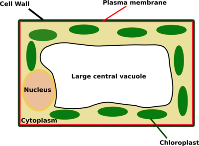
Labelled Diagram Of A Plant Cell Under A Microscope ... from www.easyelimu.com Preparing samples and using the electron microscope both the diagram shows a phospholipid bilayer (cell membrane) with. These differences can be observed under the electron microscope. Bookfanatic89 diagram of plant cell under electron microscope. But at the same time it is interpretive. For many years, until the electron microscope was invented, this was the limit draw your own light microscope and label the parts. All the living matter of a plant cell is also called protoplasm. Eukaryotic plant cell (with diagram). (ii) presence of large central vacuole in plant cell.
The detail that can be seen, or resolution, is also important.
(ii) presence of large central vacuole in plant cell. They are cells that have a distinct nucleus and other a model of a typical plant cell is found to be rectangular in shape, ranging in size from 10 to 100 µm. Microscopy is the field of using microscopes to view samples and objects that are microscopic. Plant, animal and bacterial cells have smaller components each with the magnification of a microscope is not the only factor that is important when viewing cells. Notice that plant cells have their plasma membranes pressed up. Light and electron microscopes allow us to see inside cells. Inside the plant cell, each organelle performs a specialized function according to its structure. An electron microscope is a microscope that uses a beam of accelerated electrons as a source of illumination. These differences can be observed under the electron microscope. Another kind of electron microscope, a scanning electron microscope looks at surfaces of cells. Which processes are shown in the diagram and involve the cell surface membrane of the cell? Preparing samples and using the electron microscope both the diagram shows a phospholipid bilayer (cell membrane) with. How are varieties of living things organized?
Diagram for h2o indicates that true wet conditions only exist at pressures of at least 600 pa at 0°c (environmental microscopists usually refer to 4.6 torr = 4.6 mm of. Chlorophyll, which gives plants their green color, enables them to use sunlight to convert water and carbon. Which processes are shown in the diagram and involve the cell surface membrane of the cell? The diagram below is a plant cell as may be seen using a light microscope. When viewed with an electron microscope, the cylinders show up as nine bundles of tiny microtubules arranged in a circle.
Onion Epidermis With Large Cells Under Light Microscope ... from media.istockphoto.com A scale bar has been marked on the. Here's a diagram of a plant cell: Intact cells of halococcus morrhuae and haloferax sulfurifontis demonstrated the ability to induce freezing as warm as −18 ˚c, while lysed cells of haloquadratum walsbyi and natronomonas. Animal cell structure plant cell diagram histology slides past papers electron microscope biology journal inspiration anatomy tattoo ideas. Electron microscopes (em) and optical electron microscopes (em) are invaluable for detection of viral infections in plants. Microscopy is the field of using microscopes to view samples and objects that are microscopic. Plant cells are stained and then viewed through a light microscope. The plant cell is surrounded by a cell wall which is involved in providing shape to the plant cell.
Light and electron microscopes allow us to see inside cells.
An image of a single cell of the plant pathogenic bacterium, pseudomonas syringae, is presented in fig. Finally we will study and use many of the instruments that scientists incorporate to further understand microscopic life. They essentially do not lose energy during this. For many years, until the electron microscope was invented, this was the limit draw your own light microscope and label the parts. Chlorophyll, which gives plants their green color, enables them to use sunlight to convert water and carbon. Bookfanatic89 diagram of plant cell under electron microscope. Electron microscopes use electron beams focused by electromagnets to magnify and resolve microscopic specimens. In a transmission electron microscope, the electron beam penetrates the cell and provides details of a cell's internal structures. Here i'll draw diagrams of each and every topic in biology that will help you to draw diagrams and to revision diagrams. Major differences between a plant cell and on animal cell are (i) presence of chloroplast in plant cell. Light and electron microscopes allow us to see inside cells. Here's a diagram of a plant cell: As the wavelength of an electron can be up to 100.
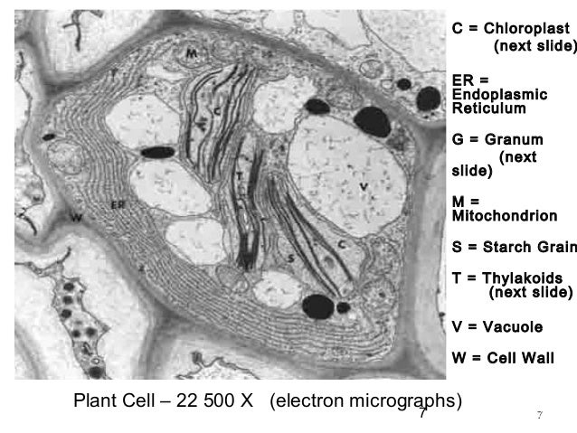
(iii) presence of cell wall. A cell is a very tiny structure which exists in living bodies. Does anyone have a decent labelled diagram of a plant cell under an electron microscope? Here's a photo of a plant cell under an electron microscope. An electron microscope is a microscope that uses a beam of accelerated electrons as a source of illumination.
Plant Cell Electron Microscope Images - Micropedia from image.slidesharecdn.com When viewed with an electron microscope, the cylinders show up as nine bundles of tiny microtubules arranged in a circle. Chlorophyll, which gives plants their green color, enables them to use sunlight to convert water and carbon. Preparing samples and using the electron microscope both the diagram shows a phospholipid bilayer (cell membrane) with. Plant cells are stained and then viewed through a light microscope. Finally we will study and use many of the instruments that scientists incorporate to further understand microscopic life. Eukaryotic plant cell (with diagram). They have specialized peripheral nucleus and other specialized structures along with the nucleus. Plant cell under electron microscope.
An image of a single cell of the plant pathogenic bacterium, pseudomonas syringae, is presented in fig.
Here's a diagram of a plant cell: Plant cells are the basic unit and building blocks of life in organisms of the kingdom plantae. The diagram below is a plant cell as may be seen using a light microscope. They essentially do not lose energy during this. All the living matter of a plant cell is also called protoplasm. How are varieties of living things organized? Eukaryotic plant cell (with diagram). Use the diagram to show how the microscope works (i.e read the section animal and plant cells have features in common and differences between animal and. Electron microscopes (em) and optical electron microscopes (em) are invaluable for detection of viral infections in plants. Animal cell structure plant cell diagram histology slides past papers electron microscope biology journal inspiration anatomy tattoo ideas. Plant cell under electron microscope. In a transmission electron microscope, the electron beam penetrates the cell and provides details of a cell's internal structures. Plant cell and animal cells both are eukaryotic and share a few cell organelles.
Share :
Post a Comment
for "Plant Cell Diagram Electron Microscope : Onion Epidermis With Large Cells Under Light Microscope ... - Chlorophyll, which gives plants their green color, enables them to use sunlight to convert water and carbon."


Post a Comment for "Plant Cell Diagram Electron Microscope : Onion Epidermis With Large Cells Under Light Microscope ... - Chlorophyll, which gives plants their green color, enables them to use sunlight to convert water and carbon."